Step 1: Scan and Model
- Step 1. Scan and Model
-
-
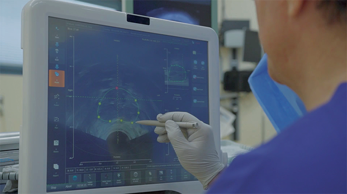
Radiologist imports MRI scan using software (UroFusion) to model prostate and mark ROI
-
Ultrasound images are automatically captured via the motorized robotic arm
-
Urologist models prostate on ultrasound images for 3D reconstruction
Step 2: Fuse and Plan
- Step 2. Fuse and Plan
-
-
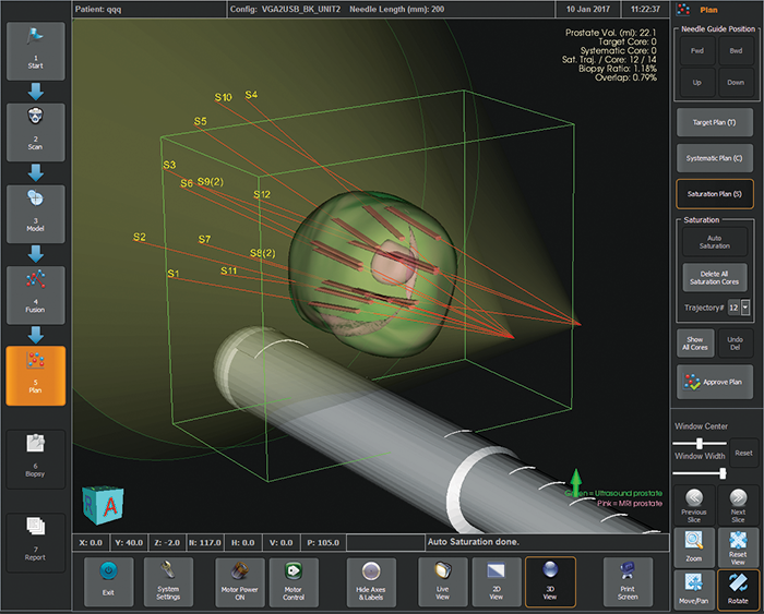
Software (UroBiopsy) instantly merges MRI and ultrasound 3D models
-
Target and saturation plans autogenerate core locations based on ROI and prostate 3D model; options for systematic plans available
Step 3: Biopsy and Report
- Step 3. Biopsy and Report
-
-
Robotic arm provides guidance for needle positioning based on physician-approved biopsy plan
-
Physician inserts the needle through the robotic guide
-
Automatic generation of comprehensive reports with clinical data and 3D images